Research:
![]() Cell-cell and cell-microenvironment interactions
Cell-cell and cell-microenvironment interactions
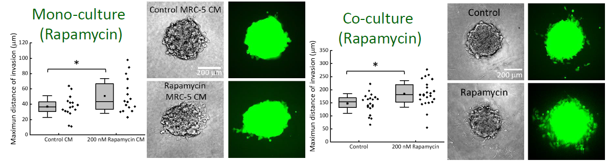
We demonstrated that the cigarette smoke extract (CSE)
treatment on fibroblast
(MRC-5) could raise the invasion capability of lung cancer cells
(CL1-0) using 3D
cellular spheroids. The cancer cells were labelled with the
CellTracker Green dye
such that we could observe and quantify the invasion distances of
individual cells
into the surrounding gel.
Fibroblast-led invasion of cancer cells from the spheroid
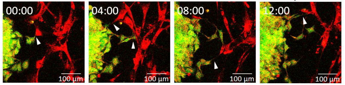
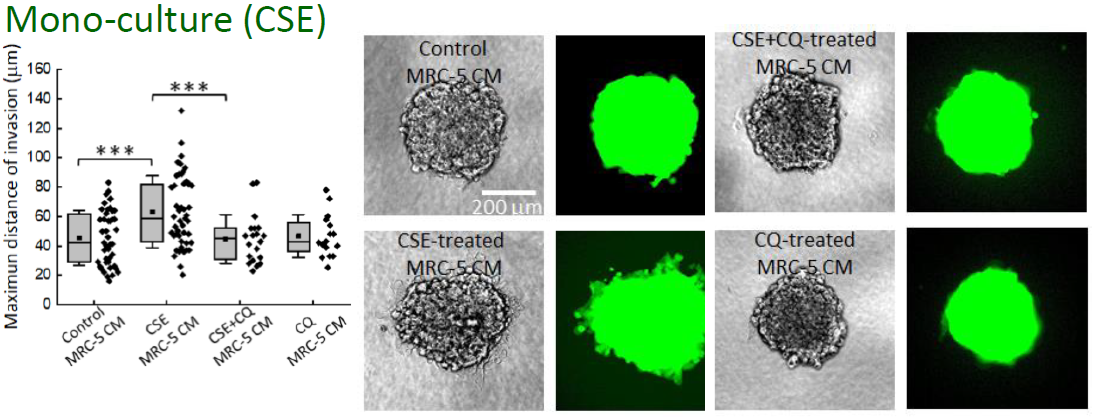
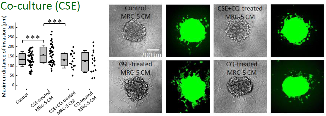
International Journal of Cancer 147, 2587 (2020).
![]() Roles of Air Gases in Lipid Bilayers
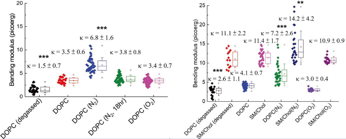
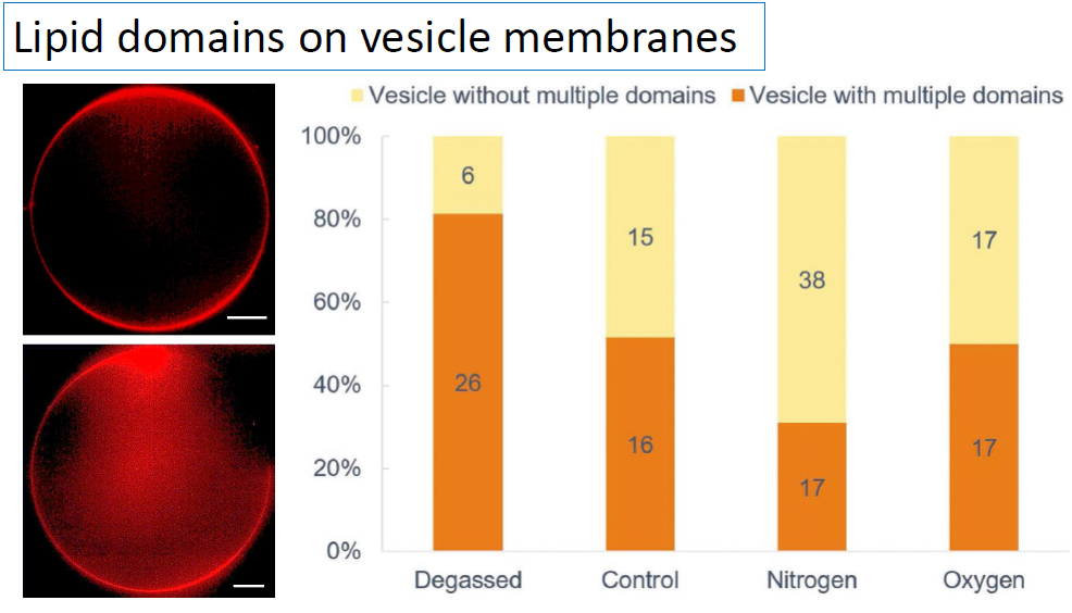
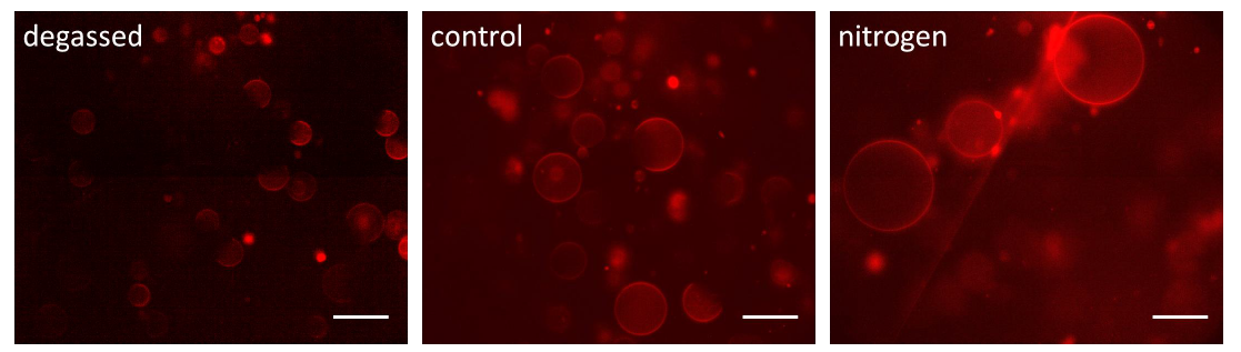
Roles of Air Gases in Lipid Bilayers
We used differential confocal microscopy (DCM) to measure the bending rigidity of vesicle membranes by quantifying membrane fluctuation amplitudes. The lipid bilayers in degassed solutions are softer and less stable than those in ambient solutions. High concentrations of nitrogen increase the bending moduli and stability of the lipid bilayers and impede domain separation in ternary lipid bilayers.
![]() Interactions between cancer cells and stromal cells
Interactions between cancer cells and stromal cells
We used microfluidic cell culture devices to investigate the communications via membrane tunneling nanotubes (TNTs) between cancer cells and myofibroblasts.
The whole device was made of PDMS by using standard soft-lithography microfabrication. More details can be found in Biomicrofluidics 8, 024107 (2014).
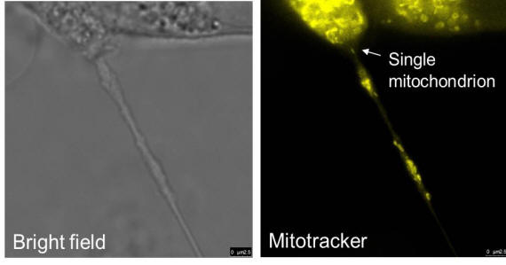
We can see mitochondria transferred through the TNT.
We used blue light (wavelength: 473 nm) to cause neurite retraction, and then a red-light (wavelength: 650 nm) spot to illuminate the soma near the junction of the retracted neurite. It seems that the red light stimulation could trigger the re-growth of a retracted neurite.
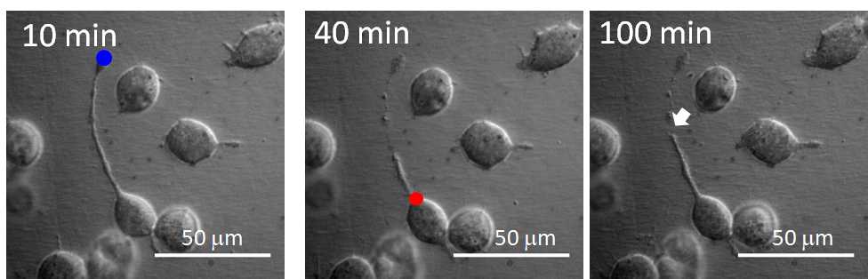
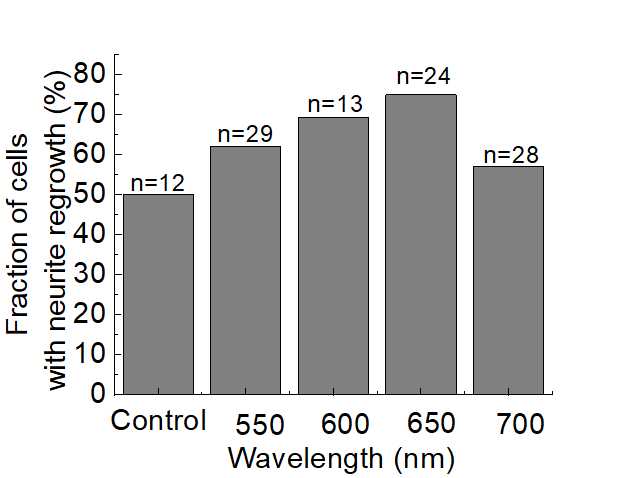
Scientific Reports 9, 18210 (2019).
![]() Cell membrane roughness and external stimulations
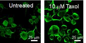
Cell membrane roughness and external stimulations
We used non-interferometric wide-field optical profilometry (NIWOP) to measure the membrane roughness on live neuroblastoma cell N2a. We found that 10 mM Taxol could reduce the membrane roughness of N2a cells. The reason could be the redistribution of microtubules caused by Taxol.
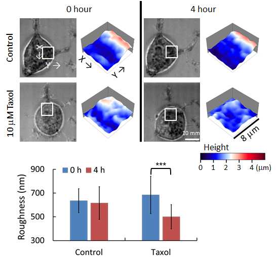
Journal of Nanobiotechnology 14, 9 (2016).
![]() SPIM observation on cellular spheroids
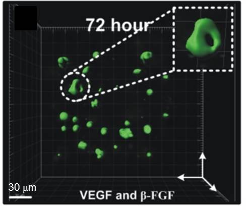
SPIM observation on cellular spheroids
In order to reduce the phototoxicity of the illumination light during 3D observation on cellular spheroids, we employed selective plane illumination microscopy (SPIM) to observe the coculture of HepG2 (red) and HUVEC (green) cells in a microfluidic device.
With the treatment of VEGF and FGF, we could see the formation of circular-cross sectional lumen structure of some HUVECs in the cellular spheroids.
Currently we are using this system to study the interaction between cancer-associated fibroblasts (CAFs) and cancer cells under various treatments.
Biomicrofluidics 8,
052109 (2014).